baby teeth skull xray
Most prematurely erupted teeth immature type are hypermobile because of limited root development. Up to 10 cash back Find the perfect baby teeth xray stock photo image vector illustration or 360 image.

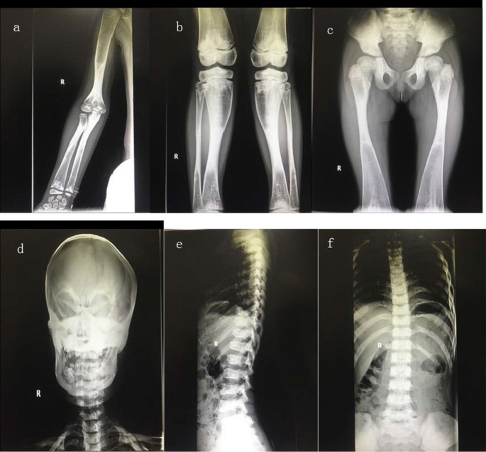
Morphologic Characteristics Of Masseter Muscle In Cleidocranial Dysplasia A Report Of 3 Cases Oral Surgery Oral Medicine Oral Pathology Oral Radiology And Endodontics
Primary baby teeth start to form between the sixth.

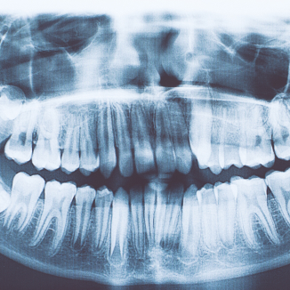
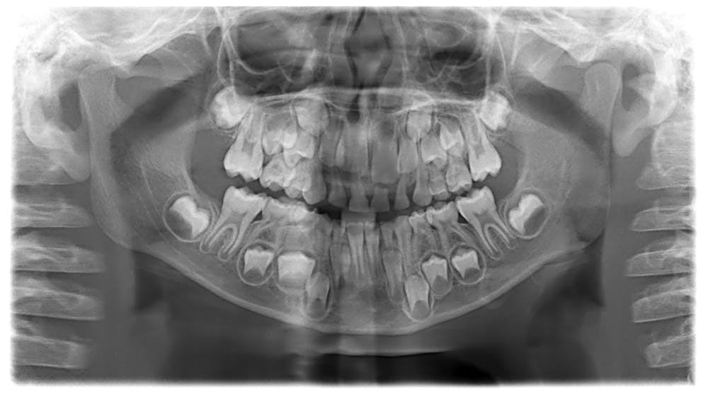
. This image is a panoramic x-ray of a child. Skull X Ray X Ray Skull Skull And Bones Pin By Leozao On De Tudo Um Pouco Video Dark Aesthetic Black Aesthetic. A skull x-ray is also used to evaluate an unusually shaped childs head.
Other conditions for which the test may be performed include. Answer 1 of 8. X-ray of a skull with surplus teeth Toddler skull X-rays are terrifying is it common.
As your childs primary teeth will not have fully. Baby teeth skull xray Monday September 19 2022 Edit. The adult teeth are only fully formed when they grow out completely however they exist in the skull in a partially formed state from a very early age probably 2-3 years old or even younger.
10 August 2021 041611 AM Sophia. While somewhat gnarly sure but its still rather fascinating to see what it looks like as permanent teeth form within the skull before pushing out baby teeth. Abnormal bone growth in the middle.
Childs skull Skull Baby teeth. Next the hard tissue that surrounds the teeth is formed around. Dental X-rays are considered a safe and effective method to gain insight into your childs oral development and health needs.
This is when the basic substance of the tooth forms. Dont front with me. Available for both RF and RM licensing.
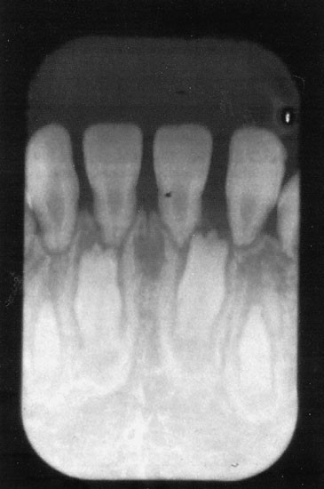
A reverse image search reveals the X-ray actually shows the skull of an 11-year-old girl with a condition called hyperdontia which is defined by the presence of supernumerary. Check out our skull teeth x ray selection for the very best in unique or custom handmade pieces from our shops. Theres a picture on Pinterest depicting a childs skull with all deciduous teeth baby teeth still attached and adult teeth showing in a quite developed stage.
Baby teeth skull xray Sunday May 8 2022 Edit. A Dental X-ray helps find problems inside the teeth such as tooth decay especially early stage decay between the teeth damage to the structure of. Tooth development happens in the womb so both babymilk and adult teeth are present when you are born.
The first stage begins in the fetus at about 6 weeks of age. 10 August 2021 040603 AM. We like to use x-ray technology to identify cavities when they are small and limited to the outer layers of the tooth before the cavity has a chance to cause any pain.
2 blood pressure 11076 heart rate 108 respirations 32 rapid and. A Child S Skull Before Losing Baby Teeth I Am Never Going Near A Child Again Www Reddit Com Childs Skull Impacted Tooth Baby Teeth X Ray Smile Young Person You Can. Stock photos 360 images vectors and.

Child Skull Memento Mutter
Dental Development After Successful Treatment Of Infantile Osteopetrosis With Bone Marrow Transplantation Bone Marrow Transplantation

Plagiocephaly Radiology Reference Article Radiopaedia Org
A Novel Mutation In Tnfrsf11a Gene Causes Pediatric Osteopetrosis Case Report Bmc Surgery Full Text

Radiology Sciencedirect
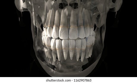
3d Rendering Teeth Skull Xray Front Stock Illustration 1162907863 Shutterstock
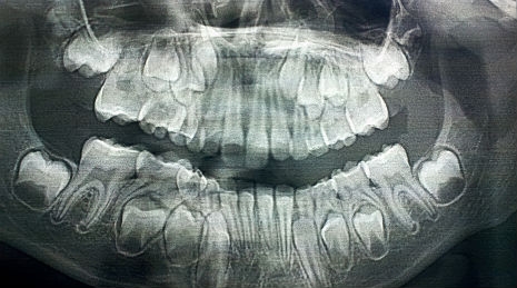
X Rays Of Kids Skulls As Their Baby Teeth Get Pushed Out And Their Permanent Teeth Grow In Dangerous Minds
Dental Technology In Modesto Ca Allure Dental Care Orthodontics

A Childs Skull Before Loosing Baby Teeth Via National Geographic Childs Skull Baby Teeth Skull

Fact Check X Ray Shown In Viral Post Is A Health Condition Not A Generic Toddler Scan Vishvas News

Do Kids Need Dental X Rays The New York Times
Differential Diagnosis Of Perinatal Hypophosphatasia Radiologic Perspectives Springerlink

Calvarial Doughnut Lesions And Osteoporosis A New Three Generation Family And Review Jaakkola 2009 American Journal Of Medical Genetics Part A Wiley Online Library

Radiology Sciencedirect

Teeth What Radiologists Should Know Radiographics
Curious Kids Why Do We Lose Our Baby Teeth

Radiology Sciencedirect

Case Series Of Cleidocranial Dysplasia Radiographic Follow Up Study Of Delayed Eruption Of Impacted Permanent Teeth

Imaging Spectrum Of Calvarial Abnormalities Radiographics